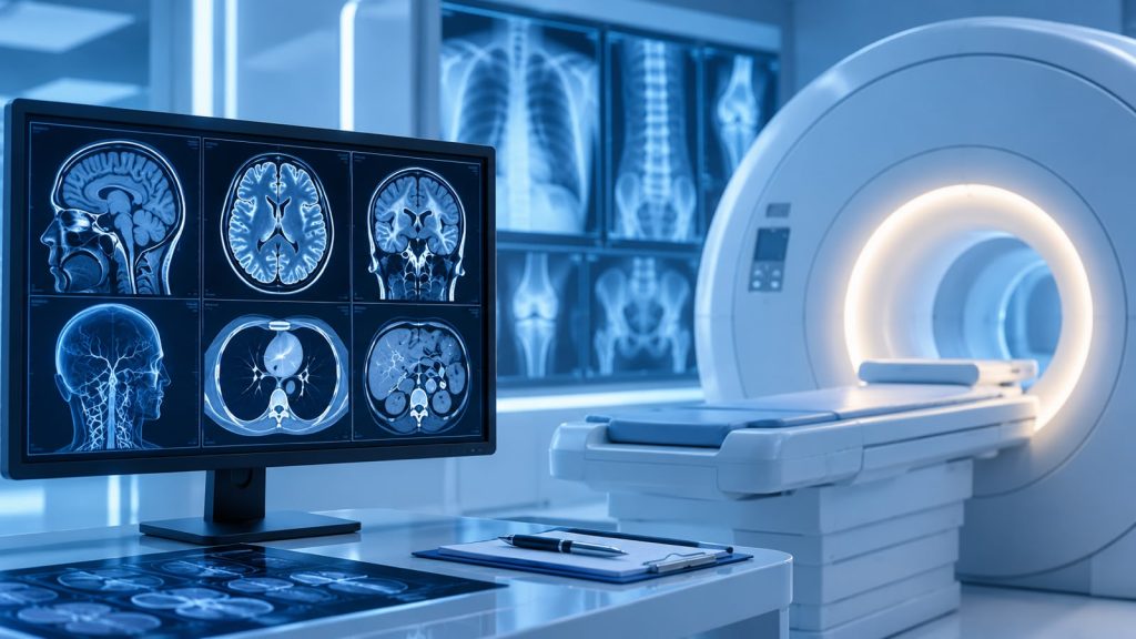
WHAT IS THE DIFFERENCE BETWEEN A CT SCAN AND MRI?

Getting a CT Scan or an MRI are on the most commonly used imaging tests in healthcare. and most of us have heard about them and know that both these scans require patients to lie on a movable table that passes through a big, donut-shaped machine. But do you know what exactly is the difference between them and how do doctors decide which one is the right choice?

Both CT scans and MRIs create detailed images of the body’s internal structures, which helps doctors  diagnosing an illness, investigating unexplained symptoms, monitoring treatment progress, or detecting conditions such as cancer. However the key difference lies in the technology used in the process. Computed Tomography scans, known as CT scans, use a series of X-rays taken rapidly from multiple angles around the body. These images are then combined to create detailed, three-dimensional views of organs and bones. On the other hand, Magnetic Resonance Imaging, i.e, MRI uses a powerful magnet and radio waves that interact with molecules in the body, producing signals that help create highly detailed images of different tissues in our body.

Doctors recommend either CT scan or MRI based on the diagnostic information required.CT scans excel at spatial resolution, meaning they are particularly effective at showing the shape, size, and boundaries of structures within the body. They help doctors clearly identify where one structure ends and another begins. MRIs, meanwhile, are known for their superior contrast resolution, allowing them to distinguish between different types of soft tissues more effectively. This makes it easier to identify subtle differences between normal and abnormal tissue.

Moreover  CT scans are often preferred when evaluating bones or when a quick assessment is required. As the scan is much faster than an MRI  it is particularly useful for patients who are in pain or may find it difficult to remain still for extended periods. MRIs often take anywhere between 30 minutes to an hour, but they can provide more detailed information about soft tissues and are frequently used when doctors need a closer look at an area of concern. 

Certain medical conditions may also influence which imaging method is chosen. For cancer diagnosis, CT scans are useful in imaging bone and air filled spaces and is often used in Lung cancer, pancreatic cancer and  bone cancer whereas MRIs provide superior soft-tissue contrast without using radiation which is useful in spotting tumours in Spinal cord and Brain, Breast cancer and Prostate cancer. 

The presence of metal implants, pacemakers, prosthetics or foreign objects embedded in your body may also influence the imaging procedure recommended by your healthcare provider. Since a very powerful magnet is involved in MRI, if the patient’s body has anything ferro-magnetic (iron-based), it could become dislodged and move around inside your body during an MRI. 

Although both scans provide valuable diagnostic information, neither is inherently better than the other. Instead, they are complementary tools that offer different types of insights.. While they differ in how they work each serves a unique purpose. Understanding these differences can help patients feel more informed and confident when undergoing medical imaging.
Read More

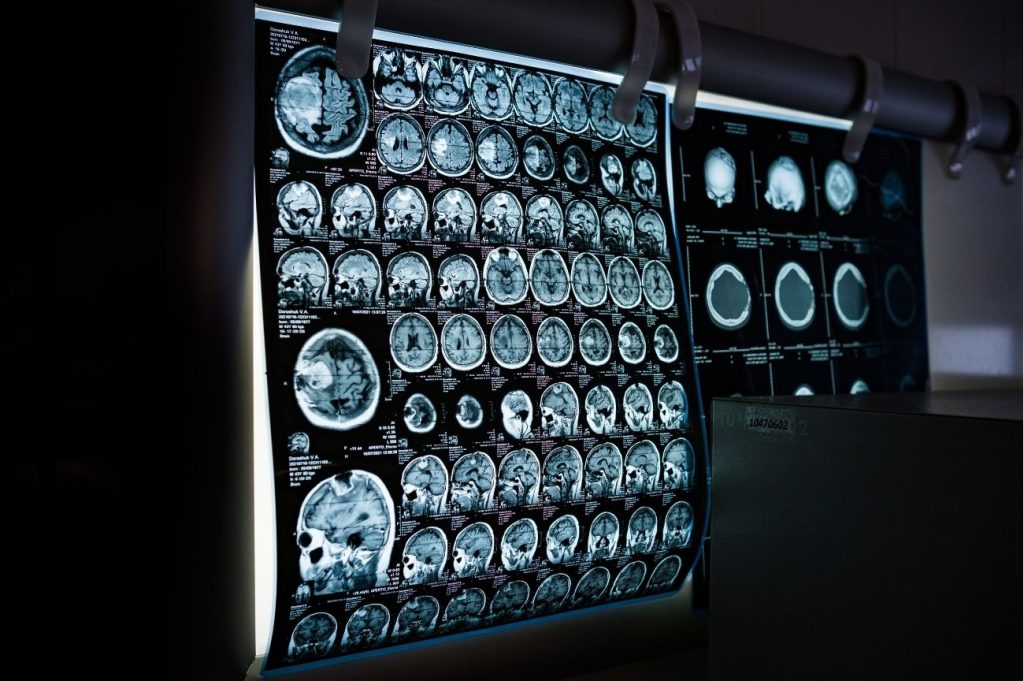
MEDICAL IMAGING IN THE AGE OF AI

Medical imaging has transformed healthcare by enabling visualization of the human body in ways that were once impossible. Technologies such as X-rays, CT scans, MRI, ultrasound, and other imaging modalities have become essential tools for diagnosing diseases, monitoring treatment, and guiding clinical decisions. As medical imaging continues to evolve it is taking a new shape with the emergence of Artificial Intelligence offering new possibilities for improving diagnostic accuracy, efficiency, and patient care.

AI-powered tools are helping healthcare professionals with the increasing volume and complexity of medical imaging data by analyzing medical images with remarkable speed and precision. In many cases, AI algorithms can identify subtle patterns and abnormalities that may be difficult to detect through visual assessment alone. This has the potential to improve diagnostic accuracy, reduce false negatives, and support earlier detection of diseases.

One of the most promising applications of AI in medical imaging is its ability to enhance efficiency. Routine and time-consuming tasks such as image segmentation, measurements, and calculations can be automated, allowing radiologists to focus more on complex cases and patient care. In ultrasound imaging, for example, AI is already being used to automate measurements such as cardiac ejection fraction and bladder volume. Advanced technologies including deep learning and convolutional neural networks have further improved the ability of AI systems to analyze medical images and support clinical decision-making.

This development is also contributing to earlier disease detection and preventive healthcare as AI has demonstrated the ability to identify signs of conditions such as cancer and cardiovascular disease at earlier stages. Early detection can enable timely intervention and improve patient outcomes. In addition, AI can support personalized medicine by analyzing medical history, imaging findings, and other patient data to help identify more targeted treatment approaches.

Despite these advantages, the integration of AI into medical imaging is not without challenges. One significant concern is bias within AI systems. Because AI models must be trained using large volumes of imaging data, the quality and diversity of those datasets directly influence performance. If the training data are biased or unrepresentative, AI systems may produce inaccurate results for certain patient populations, potentially leading to unequal healthcare outcomes.

Data privacy and security represent another important consideration. Medical imaging data contain sensitive patient information, and protecting patient confidentiality remains essential. The use of imaging data for AI development requires careful attention to consent, data protection, and privacy safeguards.

Transparency is another challenge. Many AI algorithms are highly complex, making it difficult to fully understand how they arrive at specific conclusions. This can affect trust, accountability, and adoption within clinical practice. Regulatory approval processes can also be lengthy and complex, potentially slowing the introduction of innovative AI-powered technologies into healthcare settings.

While AI has demonstrated tremendous potential, human expertise remains indispensable. AI is best viewed as a tool that supports healthcare professionals rather than replaces them. Studies have shown that the combined performance of clinicians and AI systems can achieve higher sensitivity and lower false-positive rates than either working independently. As a result, the future of medical imaging is likely to be built on collaboration between human expertise and intelligent technologies.

As AI continues to evolve, maximizing its benefits will require rigorous validation, transparent algorithms, strong ethical frameworks, secure data practices, and ongoing collaboration between technology developers and healthcare professionals. When implemented responsibly, AI has the potential to further enhance diagnostic accuracy, improve efficiency, and contribute to better healthcare outcomes for patients worldwide.

Read More

This field is for validation purposes and should be left unchanged.